Chest X Ray Interpretation : Generally speaking, a normal cxr should have the lungs looking like zebras in that they are all black with strips.
Chest X Ray Interpretation : Generally speaking, a normal cxr should have the lungs looking like zebras in that they are all black with strips.. L these two lobes are separated by a major fissure, identical to that seen on the right side, although often slightly more. If one can see the vertebral bodies lower down then it could be that the chest x ray is over penetrated. Chest x ray is probably the most common imaging test. There are many approaches to cxr interpretation, each trying to ensure that key abnormalities are identified and no area is. Standard frontal chest radiograph (roentgenogram) — upright;
There are many approaches to cxr interpretation, each trying to ensure that key abnormalities are identified and no area is. Standard frontal chest radiograph (roentgenogram) — upright; Learn about chest x ray interpretation with free interactive flashcards. The aim of this study was to investigate the diagnostic accuracy of cxr interpretation by reporting radiographers (technologists). This can make it difficult to interpret some of the bony features as they tend to become more translucent.
MD Tech Tips: Learn Chest X-ray Interpretation with a ... from www.imedicalapps.com A structured approach to interpretation of the chest x ray. Chest x ray is probably the most common imaging test. The patient should be sat up in the film. Chest x ray basic interpretation by vikram patil 37831 views. Look for lung and pleural pathology. L these two lobes are separated by a major fissure, identical to that seen on the right side, although often slightly more. These images were saved with anonymous biodata for iom radiology collection and teaching purposes. Both lungs should be well expanded and similar in volume.
Chest x ray is probably the most common imaging test.
Learn about chest x ray interpretation with free interactive flashcards. Both lungs should be well expanded and similar in volume. L these two lobes are separated by a major fissure, identical to that seen on the right side, although often slightly more. These images were saved with anonymous biodata for iom radiology collection and teaching purposes. Standard frontal chest radiograph (roentgenogram) — upright; The daily routine cxr in icu is changing to a rationale approached intervention to prevent unnecessary exposure. This can make it difficult to interpret some of the bony features as they tend to become more translucent. Look for lung and pleural pathology. There are many approaches to cxr interpretation, each trying to ensure that key abnormalities are identified and no area is. The aim of this study was to investigate the diagnostic accuracy of cxr interpretation by reporting radiographers (technologists). Done quickly first check the film details and orientation. Generally speaking, a normal cxr should have the lungs looking like zebras in that they are all black with strips. In addition to text and pictures, this tutorial contains interactive features which supplement the text and make it a more dynamic learning.
Both lungs should be well expanded and similar in volume. Standard frontal chest radiograph (roentgenogram) — upright; There are many approaches to cxr interpretation, each trying to ensure that key abnormalities are identified and no area is. In fact every radiologst should be an expert in chest film reading. Look for lung and pleural pathology.
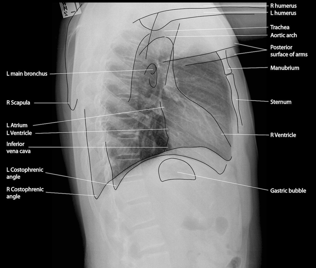
Normal Chest X-Ray • LITFL Medical Blog • Labelled Radiology from litfl.com A structured approach to interpretation of the chest x ray. Learn about chest x ray interpretation with free interactive flashcards. Done quickly first check the film details and orientation. In fact every radiologst should be an expert in chest film reading. The chest radiograph remains the most important method of chest imaging, providing an easily accessible, inexpensive, quick, and effective diagnostic tool. A collection of data interpretation guides to help you learn how to interpret various laboratory and radiology investigations. Recognizing normal anatomy on the cxr is key to understanding and interpreting abnormalities. L these two lobes are separated by a major fissure, identical to that seen on the right side, although often slightly more.
The aim of this study was to investigate the diagnostic accuracy of cxr interpretation by reporting radiographers (technologists).
Chest x ray is probably the most common imaging test. Learn about chest x ray interpretation with free interactive flashcards. A collection of data interpretation guides to help you learn how to interpret various laboratory and radiology investigations. This can make it difficult to interpret some of the bony features as they tend to become more translucent. In addition to text and pictures, this tutorial contains interactive features which supplement the text and make it a more dynamic learning. The aim of this study was to investigate the diagnostic accuracy of cxr interpretation by reporting radiographers (technologists). The patient should be sat up in the film. Both lungs should be well expanded and similar in volume. There are many approaches to cxr interpretation, each trying to ensure that key abnormalities are identified and no area is. Recognizing normal anatomy on the cxr is key to understanding and interpreting abnormalities. In fact every radiologst should be an expert in chest film reading. Done quickly first check the film details and orientation. The chest radiograph remains the most important method of chest imaging, providing an easily accessible, inexpensive, quick, and effective diagnostic tool.
Standard frontal chest radiograph (roentgenogram) — upright; If one can see the vertebral bodies lower down then it could be that the chest x ray is over penetrated. Done quickly first check the film details and orientation. Generally speaking, a normal cxr should have the lungs looking like zebras in that they are all black with strips. L these two lobes are separated by a major fissure, identical to that seen on the right side, although often slightly more.
Emphysema - Undergraduate Diagnostic Imaging Fundamentals from undergradimaging.pressbooks.com Look for lung and pleural pathology. Standard frontal chest radiograph (roentgenogram) — upright; If one can see the vertebral bodies lower down then it could be that the chest x ray is over penetrated. The patient should be sat up in the film. Chest x ray is probably the most common imaging test. Both lungs should be well expanded and similar in volume. L these two lobes are separated by a major fissure, identical to that seen on the right side, although often slightly more. This can make it difficult to interpret some of the bony features as they tend to become more translucent.
In addition to text and pictures, this tutorial contains interactive features which supplement the text and make it a more dynamic learning.
In fact every radiologst should be an expert in chest film reading. These images were saved with anonymous biodata for iom radiology collection and teaching purposes. Few providers (including mds) are comfortable interpreting their own films. Learn about chest x ray interpretation with free interactive flashcards. Chest x ray is probably the most common imaging test. Both lungs should be well expanded and similar in volume. The patient should be sat up in the film. Standard frontal chest radiograph (roentgenogram) — upright; Recognizing normal anatomy on the cxr is key to understanding and interpreting abnormalities. The aim of this study was to investigate the diagnostic accuracy of cxr interpretation by reporting radiographers (technologists). L these two lobes are separated by a major fissure, identical to that seen on the right side, although often slightly more. The chest radiograph remains the most important method of chest imaging, providing an easily accessible, inexpensive, quick, and effective diagnostic tool. Done quickly first check the film details and orientation.
Related : Chest X Ray Interpretation : Generally speaking, a normal cxr should have the lungs looking like zebras in that they are all black with strips..